Understanding Lumps and Bumps in dogs and cats

Causes, symptoms, and treatment

Key symptomsVisible lump or swelling under/above skin, may grow, change shape, or ooze; sometimes painful or ulcerated; may cause licking or irritation.
CommonalityVery common in dogs (especially older or overweight), less common in cats.
TransmissionNot contagious
Impact on healthMild to Severe –
depends on lump type (benign vs malignant)
Treatment cost~$300 to $3,000 AUD (biopsy or removal); complex cancer cases can exceed $7,000 AUD
Treatment duration2 weeks to several months
(depending on surgery and recovery)
Ease of preventionDifficult

Most affected dog breeds

Most affected cat breeds

Complete Guide to Lumps in Pets

Discovering a lump on your pet can be unsettling, but many are harmless. This guide explains what lumps are, when to seek help. And how Petsy Pet Insurance may support your pet’s care, depending on your policy.

What exactly are lumps and why do they appear?

Lumps can vary in their size, texture and location, and their causes can range from harmless fat deposits to cancerous tumours. Alongside this, some lumps can develop slowly over time or 
suddenly appear.

Lumps in both dogs and cats can occur due to:

Lumps under our policy refers to: the protuberance or localised area of swelling or unusual growth that can occur anywhere on or in the body including tumours, warts, cysts, growths, mucoceles, haematomas and abscesses

Understanding Lumps on your pet

Before diving into specific types, here are some of the most common lumps pet parents search for:

  • Soft, squishy lump under the skin – Likely a fatty lump (lipoma), which is usually harmless but should still be checked.
  • Small, raised bump on the skin – Could be a wart, insect bite, or tumor; a vet can help determine what it is.
  • Large, fluid-filled lump – May be a cyst or abscess, which can range from minor to serious.
  • Hard, immovable lump – These may be more concerning, as they can be attached to deeper tissues and require further testing.
  • Rapidly growing lump – Needs urgent vet attention, as it may be cancerous.

If you notice a lump on your pet, it’s always best to have your vet check it to ensure it’s nothing serious.

Understanding Benign vs. Malignant Lumps

Knowing whether a lump is benign or malignant helps determine the risk it poses to your pet’s health.

The only way to confirm whether a lump is benign or malignant is through a veterinary evaluation, which may include tests like fine needle aspiration, biopsy, or imaging. Early detection is key to successful treatment.

Why this matters for pet owners

Finding a lump on your pet can be worrying, but early detection and prompt veterinary care can make a big difference. Some breeds are more prone to certain types of lumps, so knowing what to look for and acting quickly can improve treatment options, including medication, surgery, or other therapies.

How are Lumps diagnosed?

Diagnosing lumps in dogs and cats starts with a vet check-up, followed by tests to determine what type of lump it is.

Physical examination

Your vet will assess the size, location, texture, and mobility of the lump to get an idea of what it might be.

Fine Needle Aspiration (FNA)

If the lump is easy to access, your vet may insert a small needle to collect cells or fluid for microscopic evaluation. This can help determine if the lump is benign or malignant.

Biopsy

If more information is needed, a biopsy may be done to take a larger tissue sample for testing.

Imaging Tests

X-rays, ultrasound, or CT scans may be used to check if the lump is deep under the skin or has spread to other areas.

Lab Testing

If needed, a sample of the lump can be sent to a pathologist for a detailed evaluation, especially if surgery is required to remove it.

These tests help your vet identify the lump and create a treatment plan tailored to your pet’s needs. If you notice a lump on your pet, early diagnosis is key to ensuring the best possible outcome.

Why an accurate diagnosis matters

Getting the right diagnosis for a lump is crucial to ensuring the best outcome for your pet. A misdiagnosis or delay can lead to unnecessary treatments or missed opportunities for early care. By identifying the type of lump and how it behaves, vets can create a targeted treatment plan that’s both effective and minimizes risk. A clear diagnosis also helps you make informed decisions about your pet’s health with confidence.

Treatment options for Lumps

Treatment depends on the size, location, and type of lump. Your vet will guide you through the best approach based on your pet’s needs.

Surgical removal

Radiation therapy

Chemotherapy

Medication & supportive care

What happens after treatment?

Your vet may recommend regular check-ups and monitoring to catch any changes early. This may include follow-up imaging or small tests on the surgical area.

Deciding on treatment can feel overwhelming, but your vet will guide you through the process to ensure your pet gets the best possible care. Pet insurance can also help ease financial concerns, so you can focus on your pet’s health and quality of life.

If you notice a lump on your pet, early diagnosis and treatment make all the difference. Reach out to your vet if you have any concerns.

Cost of diagnosis and treatment

The cost of treating lumps in pets can vary significantly depending on factors such as the type of lump, the diagnostic tests required, and the complexity of the treatment course. For instance, fine needle aspiration, can be done during a consult, and (excluding cost of consult) may range from $100 for FNA, around $500 for imaging, to over $2500 for surgical excision, depending on the location. Costs vary by location, clinic, and case complexity. Consult your Vet for exact pricing.

When to see a vet

Knowing when to consult your veterinarian is crucial. Schedule a visit if:

  • You notice single or multiple lumps appearing over a short period of time
  • The lump grows rapidly or changes in appearance (e.g. colour, texture, or size)
  • It causes obvious discomfort to your pet
  • There are signs of infection, such as redness, swelling, odour, or discharge
  • The lump is hard, immoveable, or feels deeper than just under the skin
  • Your pet shows other symptoms, such as lethargy, loss of appetite, diarrhoea, or weight loss

Your veterinarian may perform a fine needle aspiration, biopsy, or imaging to diagnose the lump and recommend the best course of action from there. Having a pet insurance policy in place can also help cover vet costs, allowing you to focus on your pet’s health with confidence.

Common types of Lumps in pets

Here are some of the most common lumps found in dogs and cats. While this isn’t a complete list, these are the ones pet parents see most often. If you notice a lump that isn’t listed or are unsure about one on your pet, it’s always best to check with your vet.

Dogs
Tumour Description Risks Treatment
Benign
Lipomas Soft, squishy, and moveable lumps just under the skin, fat deposits. Usually remain benign, however large lipomas can impede mobility and cause discomfort. No treatment is usually required. However, if they grow significantly, surgical removal may be necessary.
Fibromas Usually isolated, hairless lumps found on the head and legs. Can feel firm/rubbery or soft/squishy. Are growths of fibrous tissue such as muscle, blood vessels, fat. Usually remain benign. Only requires surgical removal if it grows larger or changes appearance.
Sebaceous Cysts Small, fluid-filled sacs formed from clogged hair follicles. Can rupture and become infected. May drain naturally or require surgical removal if it becomes problematic.
Warts & Skin Tags Often found in older dogs, can be single or multiple. Usually harmless but can become irritated. Rarely requires removal unless obvious signs of infection.
Haematomas Blood-filled swellings caused by trauma, often seen on the ears. Painful, but non-threatening. Draining or surgical correction. May be followed with an antibiotic course if infected.
Melanomas Usually found on the head and forelimbs, can either be flattened or raised, isolated or multiple darker lumps. Common in breeds such as miniature and standard Schnauzers, Doberman Pinschers and Golden Retrievers. Usually benign, there is a malignant form however it is uncommon (these would appear as raised, ulcerated lumps). Surgical removal
Malignant
Mast Cell Tumours Most common skin tumour in dogs, can occur anywhere, can look like a generic lump or progress to ulcerated masses. Can look like a lipoma. Can begin with rash-like signs and progress to vomiting, diarrhoea (high-grade tumours) Surgical removal and radiation or chemotherapy, based on degree.
Soft Tissue Sarcomas Lumps are often found on paws, mouth, or skin, but can occur anywhere with connective/’soft tissues’ such as muscles, neural tissues, fibrous tissues. Can appear as isolated or multiple, firm, deep, and fleshy-feeling lumps. Often requires diagnostic tools to determine if it is malignant. If it spreads to internal organs, particularly the lungs, it can be fatal. High rate of recurrence: Even if one lump is removed, there is a highly likely chance that another will occur. Surgical removal and radiation, or chemotherapy.
Squamous Cell Carcinomas (SCC) Most frequently diagnosed skin carcinoma in dogs. Two forms:
  1. Skin: Firm, raised, often ulcerated that appear on the head, lower legs, abdomen, and rear.
  2. Subungual: Originate from under the nail/claw. Frequently associated with dark-haired breeds
Are often solitary, however multiple can develop, especially in areas with increased sun exposure. The lumps due to the skin form of SCC are sometimes cause by sun exposure, especially in white-skinned, shorthaired breeds. Before the malignant tumour develops, they will first have solar keratosis; Solar keratosis is distinguished by thickened and discoloured skin and should be checked by a veterinarian. Can often return around 20 weeks post-surgical removal. Surgical removal, including amputation of an involved toe or ear. Combined with radiation treatment or chemotherapy. Prevention: reduce exposure to excessive sunlight.
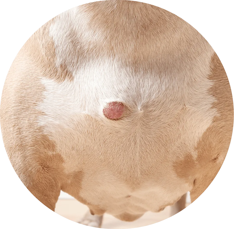
Cutaneous/ Subcutaneous Haemangiosarcoma Can appear as a red or purple lump or nodule, bruising or bleeding around the mass. Other symptoms can include lethargy, lameness, loss of appetite or weakness. When cutaneous haemangiosarcoma is caught early, these dogs typically do well. However, there are other forms of haemangiosarcoma (subcutaneous, splenic and cardiac) that are much more aggressive and carry a less promising prognosis. Surgical removal. Other medications may also be administered such as anti-bleeding medications or blood transfusions in severe cases.
Anal Sac Tumours Lumps appear as firm, deeper, masses underneath the rectum, where the anal sacs are located. As tumours grow, it can compress the rectum and induce constipation. High rate of spread to other organs, can be fatal. Surgical removal or reduction of anal sacs, alongside with radiation and/or chemotherapy.
Cats
Tumour Description Risks Treatment
Benign
Lipomas Rare in cats but also seen as soft, squishy, and moveable lumps just under the skin. Usually remain benign, however large lipomas can impede mobility and cause discomfort. No treatment is usually required. However, if they grow significantly, surgical removal may be necessary.
Fibromas Similar to dogs, appear as isolated, raised, hairless lumps that can either feel firm/rubbery or soft/squishy. They are growths of fibrous tissue such as muscle, blood vessels, fat. Usually remain benign but can grow and impede mobility and cause discomfort. There is a malignant form, refer below to ‘Fibrosarcomas’. No treatment is usually required. However, if they grow significantly, surgical removal may be necessary.
Keratinised Skin Cysts Can be solitary or multiple lumps, that are firm and hardened, and usually the same colour as the hair. These lumps can be variable in size and are full of keratin. If squeezed, can release keratin into adjacent areas and cause inflammation Surgical removal
Feline acne Small bumps that typically develop on the chin, lips, and/or face. Usually remain benign and can appear dirty. Can become swollen and inflamed. If it becomes a chronic issue, harder, crusty, and painful lumps may develop. Improved hygiene: may include antibacterial shampoos, prescribed antibiotics (ointment or oral) from your veterinarian or oral antibiotics/steroids in more chronic cases.
Basal Cell Tumour Basal cells are the topmost layer of the skin. Common in older cats and longhair breeds. Appear as isolated, firm, and hairless lumps. Can be ulcerated. Variable in size (between 1-10cm) and often darker in colour Cysts can result from basal cell tumours. There is also a malignant form, refer bellow to ‘Basal Cell Carcinoma’. Surgical removal
Mast Cell Tumours Uncommon in cats, usually found on the head and neck. Two forms:
  1. Mast cell type (more common): Appear as isolated, hairless lumps that range between 2-3cm.
  2. Histiocytic type: most common in Siamese cats <4 years old. Appear as multiple, small, firm lumps.
If lumps on the skin are found to be mast cell tumours, usually benign. But if lumps are found attached to internal organs using diagnostic imaging, poorer prognosis. Surgical removal (mast cell type) or no treatment (histiocytic type)
Malignant
Lymphoma One of the most common malignant tumours in cats, gastrointestinal form of lymphoma is the most common. Lumps can be found on the skin after the original tumour has spread. If it originates from the skin, known as epitheliotropic skin lymphosarcoma, lumps can appear as itchy, crusty plaques. High rate of spread to other internal organs/areas of the body – these forms are much more aggressive cancers. High rate of recurrence: Even if one lump is removed, there is a highly likely chance that another will occur. Dependent on the type of lymphoma, treatment can range from palliative care to surgical excision and/or radiation or chemotherapy
Fibrosarcoma Common soft tissue tumour in cats. Can appear lumpy, deep, and firm under the skin. High rate of recurrence and spread. Surgical removal followed with radiation or chemotherapy
Basal Cell Carcinomas Can appear as ulcers on head, legs, and/or neck. Not usually raised masses as seen in Basal Cell Tumours. Can spread to other areas of the skin, causing more ulcers to form. Surgical removal
Feline Injection Site Sarcoma Uncommon. Variably sized lumps found on the back of the neck to shoulder region (where an injection would normally go). Can develop years after injection occurred. Are highly aggressive tumours, requires advanced imaging pre-surgically to determine margins. Surgical removal

Prevention Tips (where applicable) for Lumps

While many lumps cannot be prevented, here are a few steps that can be taken to monitor and prevent when possible:

Regular grooming & skin checks

Inspect your pet’s skin during grooming or cuddle sessions to catch lumps early.

Healthy diet & exercise

Maintaining a healthy weight can lower the risk of lipomas and other health issues, such as joint disease.

Parasite prevention

Use flea, tick, and mosquito preventatives to reduce the risk of bites and 
related lumps.

Timely vaccinations

Some lumps, such as post-vaccination nodules, are normal but ensuring proper vaccination schedules is crucial for the overall wellbeing and health of your pet. 

Getting Lumps covered with pet insurance

Does Petsy cover Lumps?

Yes, as long as they are not pre-existing and your pet is outside their exclusion periods.

If your pet was under 1 year old when you started the policy, lumps have a 14-day exclusion period.

If your pet was 1 year or older, a 6-month exclusion period applies.

What is a pre-existing symptom or condition?

A pre-existing condition is any injury, illness, or condition (including lumps) that:

  • Showed signs or symptoms before your policy started or during the 
exclusion period.
  • Was diagnosed, treated, or investigated by a vet before coverage began.
  • Was something you were aware of (or reasonably could have been aware of) before coverage started.

If a lump appeared before your exclusion period ended, it would be considered pre-existing and not covered under your policy.

Understanding pre-existing conditions for Lumps

Whether a lump is considered a pre-existing condition depends on when it first appeared in relation to your policy’s exclusion periods. Here are some examples:

When you start your policy along with your vet’s clinical notes are key in determining if a lump is considered pre-existing. Since lumps are also classified as a Specified Condition, they’re subject to a 6-month exclusion period unless a waiver form is completed.

If you’re unsure whether a lump would be covered, we recommend speaking with your vet or requesting a pre-existing condition assessment.

How Petsy Pet Insurance can help with Lumps

  • If the lump is not pre-existing and you’re past the exclusion period, we’ll be able to help cover the costs of treating your pet.
  • If your pet is 1 year or older, lumps have a 6-month exclusion period, but you can apply to reduce this by completing a Waiver Form.
  • If your pet was less than 1 year when applying for a new policy, the exclusion period for lumps is 14 days.

How do I apply to reduce the 6 month exclusion period?

  • Print the waiver form and take it to your vet.
  • Ask your vet to examine your pet and complete Part 2 of the form.
  • Both you and your vet must sign the form.
  • Return the completed form to us within 14 days of your vet visit.
  • Once reviewed, we’ll send you an updated Certificate of Insurance reflecting any reduced exclusion periods.

If you’ve recently found a lump on your pet, we recommend speaking to your vet as soon as possible. Early detection can make a big difference in treatment options and outcomes. If you have any questions, our team is always here to help.

Looking after your
pet’s health

Finding a lump on your pet can be concerning, but remember that many lumps are harmless. Knowing the difference between benign and malignant lumps, understanding treatment options, and keeping an eye on any changes can help you feel more confident in managing your pet’s health.

Thanks to modern veterinary care, there are many options available, from simple procedures for benign lumps to more advanced treatments for serious conditions. Pet insurance can also help ease financial worries, so you can focus on what matters most—your pet’s well-being.

Regular grooming and skin checks can help you spot new lumps early, and keeping an open dialogue with your vet ensures your pet gets the care they need. Whether it’s monitoring, treatment, or supportive care, your vet is there to guide you.

By staying informed and proactive, you can help your pet live a happy, healthy, and comfortable life.

Specified Conditions includes:

  • Cruciate ligament damage
  • Intervertebral disc disease
  • Hip dysplasia
  • Patella luxation
  • Elbow dysplasia
  • Cherry eye
  • Entropion
  • Ectropion,
  • Osteochondritis dissecans (‘OCD’)
  • Lumps

Lumps means the protuberance or localised area of swelling or unusual growth that can occur anywhere on or in the body including tumours, warts, cysts, growths, mucoceles, haematomas and abscesses.

You can apply to reduce the 6-month exclusion period for Specified Conditions by using our request for Waiver Form. Simply take this form to your next vet visit and submit the completed form back to [email protected] within 14 days of the visit.

Optional Extra Benefits

During the application process You will be provided with the option to include Optional Extra Benefits that cover certain conditions and Treatments which are not otherwise covered under the Policy.

The Optional Extra Benefits are:

Alternative Therapies, Behavioural Problems, and Dental Illness.

Examples of Alternative Therapies: Acupuncture, physiotherapy, hydrotherapy.

Examples of Behavioural Problems: Excessive licking, fur pulling, pacing and destructive chewing.

Examples of Dental Illnesses: Dental diseases, gingivitis, periodontal disease.